Uterosacropexy by Dr. sushila saini
Hellow guys, Welcome to my website, and you are watching Uterosacropexy by Dr. sushila saini. and this vIdeo is uploaded by JaipurDoorbeen Hospital at 2017-08-04T04:51:57-07:00. We are pramote this video only for entertainment and educational perpose only. So, I hop you like our website.
Info About This Video
| Name |
Uterosacropexy by Dr. sushila saini |
| Video Uploader |
Video From JaipurDoorbeen Hospital |
| Upload Date |
This Video Uploaded At 04-08-2017 11:51:57 |
| Video Discription |
A sacrocolpopexy is a surgical procedure used to treat pelvic organ prolapse. Pelvic organ prolapse is a condition that is caused by a weakening of the normal support of the pelvic floor, and is similar to a hernia in the vagina.
Laparoscopic Sacro colpopexy [LSCP] is the gold standard procedure for pelvic organ prolapse repair since long. This approach is justified in terms of correct anatomical location. This procedure can be performed open or laparoscopic.
This approach was first published in 1920 but not in full swing till 1960s when artificial tissue like mesh came into play to bridge the gap between vault/cervix to the sacrum.
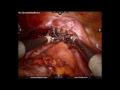
In This technique, a Y-shaped mesh placed posteriorly and anteriorly to the vaginal wall and sutured in conjunction to the apical structure (cervix and vault), and the distal part of the mesh is anchored to the anterior longitudinal ligament on promontory or sacrum.
Steps of surgery:
1. Placement of trocars
2. Opening the peritoneum over the promontory
3. J cut in right pelvic peritoneum till the cervix, medial to uterosacral ligament [USL]
4. Opening the pod
5. Dissection between the rectum and the vagina till the levator ani muscle
6. Opening the cervico-vesicle peritoneum and separation of bladder
7. Making opening in broad ligament
8. Placement of posterior mesh
9. Placement of anterior mesh
10. Closure of peritoneum over the mesh
After examining pelvic structures, lift peritoneum over sacral promontory and give a nick with scissors/ harmonic/bipolar shearer. Co2 enters into the space and helps in dissection. Extend this cut into right lateral pelvic wall, medial to uterosacral ligament and lateral to rectum into pararectal space.
Inferior hypogastric nerve found in this space only adhered to rectum in fatty tissue so keep an eye while dissecting the sub peritoneal fat in pararectal space.
Don’t dissect lateral to USL, as it may damage ureter present just lateral to USL.
Now make a J shape curve at base of uterosacral ligament towards POD till the opposite uterosacral ligament.
After reaching at POD, dissect between two layers of Denonvilliers fascia so that rectum and vagina.
Now start dissecting between the rectum and vagina till the pelvic floor or levator ani muscle seen or as low as possible to anchor pelvic floor or perineal body with mesh and to the promontory, to give complete repair of pelvic floor descent.
Next step to identify cervico-vesicle fascia and lift it up with grasper and cut with scissor to separate bladder from cervix with sharp or blunt dissection.
Make hole in broad ligament at level of isthmus to pass the mesh around the cervix to hold it.
After completing the dissection both in anterior and posterior compartment, prepare the mesh.
A macroporous mesh of 15*15cm is good enough.
Macroporous mesh has the advantage of less burden of mesh with larger pore size for easy movement of fibroblasts to grow between into interstices.
You may use single strip of mesh broad at pelvic floor end and narrow at top end towards sacral promontory or
you may design design the mesh into “clover leaf pattern” shape to place the one broad arm to the levator ani muscle over the rectum, opposite end goes to promontory and lateral arm encircle the cervix, through the opening made into broad ligament, to fix anteriorly. or
you may take One more strip of mesh to wrap around the cervix and fix it with 2-0 ethibond suture to the cervix anteriorly and to the USL & mesh posteriorly. This strip of mesh may have central tongue like projection of 3-4 cm which will be set down below the bladder and fixed with bladder pillars and cervix to take care of cystocele in case of anterior compartment defects like we take in Pectopexy.
To fix the mesh with absorbable tacks on pelvic floor as suturing may be difficult to deep into the pelvis or you can use 2-0 ethibond suture on 26mm half circle needle to fix the mesh.
To fix the mesh, use always permanent suture.
Now fix the uterus over the mesh after pulling inside the pelvis to keep it on normal position. Take suture from uterosacral ligament and posterior wall of cervix and then into the posterior mesh which is already fixed over levator ani.
Now prepare the promontory for mesh fixation – after opening the peritoneum, sub peritoneal fat need to dissect off from anterior longitudinal ligament [ALL] to expose it fully.
Pull the end of the posterior mesh to the promontory and fix it here with the help of permanent titanium tacks or sutures, both options are viable. Using tackers is an easier option but costly.
Now close peritoneum over the mesh to reperitonise it. |
| Category |
Education |
| Tags |
laparoscopic sacrocolpopexy | sacrocolpopexy | uterosacropexy | sacropexy | uteropexy | prolapse surgery for uterine prolapse | best surgery for uterine prolapse | how to preserve uterus in prolapse | uterus sparing surgery for uterine prolpase |
More Videos