Endoscopic sinus surgery: Anterior ethmoidectomy dissection.
Hellow guys, Welcome to my website, and you are watching Endoscopic sinus surgery: Anterior ethmoidectomy dissection.. and this vIdeo is uploaded by Dr Ricardo Bartel at 2020-09-24T09:29:27-07:00. We are pramote this video only for entertainment and educational perpose only. So, I hop you like our website.
Info About This Video
| Name |
Endoscopic sinus surgery: Anterior ethmoidectomy dissection. |
| Video Uploader |
Video From Dr Ricardo Bartel |
| Upload Date |
This Video Uploaded At 24-09-2020 16:29:27 |
| Video Discription |
A short demonstration of an anterior ethmoidectomy dissection.
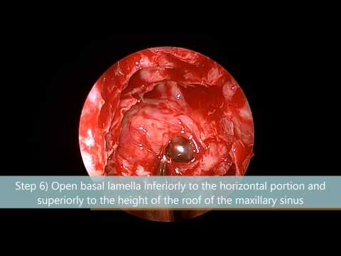
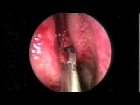
The bulla ethmoidalis is a constant, variably sized landmark and is the entry point into the anterior ethmoid complex. It is safely entered infero-medially using any kind of instrument. Bone and sinus mucosa are removed, and dissection proceeds on a broad front; the next ethmoid air cell is then identified and entered. The lateral limit of the dissection is the lamina papyracea, the medial limit the middle turbinate, the superior limit the skull base, and the posterior limit the basal lamella, which is part of the attachment of the middle turbinate to the lateral nasal wall and separates the anterior and posterior ethmoid air cells.
The key structures at risk during this step of the surgery are the orbit and the skull base. Staying medially to a sagittal plane, defined by the medial wall of the maxillary sinus, ensures that the surgeon does not enter the orbit, assuming that the medial wall of the maxillary sinus has not been lateralized (as in silent sinus syndrome). Or the medial wall of the orbit is not medialized (as in previous orbital wall decompression). The risk of accidental entry into the orbit is increased if an infraorbital cell is present, which is in 12% of patients. In 8% of patients, the cribriform plate and the lateral lamella are low-lying and asymmetrical. This increases the risk of skull base and intracranial complications. The risk of orbital injury further increases when the supraorbital region is pneumatized. Supraorbital pneumatization is also usually associated with low-lying "hanging" anterior ethmoid arteries, which could be damaged during the anterior ethmoid dissection and should be identified preoperatively.
All the above emphasizes the importance of careful analysis of CT scans prior to surgery. It is important to avoid instrumentation of the frontal recess during this phase of surgery. In most cases, complete clearance of the anterior ethmoids followed by adequate medical treatment is sufficient for the treatment of frontal sinus disease. One should always remember that instrumentation in the frontal sinus greatly increases the risk of iatrogenic stenosis and difficult to treat frontal sinus disease.
#doctor #medicine #medicina #surgery #cirugia #surgeon #cirujano #otolaryngology #ent #otolaryngologist #otorrinolaringologia #orl #otorrino #endoscopy #endoscopia #anatomy #anatomia #meded #medicaleducation #otology #rhinology #endoscopicsinussurgery #endoscopicearsurgery #sinusitis #otitis |
| Category |
Science & Technology |
| Tags |
Science & Technology Download MP4 | Science & Technology Download MP3 | Science & Technology Download MP4 360p | Science & Technology Download MP4 480p | Science & Technology Download MP4 720p | Science & Technology Download MP4 1080p |
More Videos