Early Surgical Exploration of Traumatic Frontal (Temporal) Branch Facial Nerve Paralysis
Hellow guys, Welcome to my website, and you are watching Early Surgical Exploration of Traumatic Frontal (Temporal) Branch Facial Nerve Paralysis. and this vIdeo is uploaded by Dr Thomas McClellan at 2015-07-30T11:42:27-07:00. We are pramote this video only for entertainment and educational perpose only. So, I hop you like our website.
Info About This Video
| Name |
Early Surgical Exploration of Traumatic Frontal (Temporal) Branch Facial Nerve Paralysis |
| Video Uploader |
Video From Dr Thomas McClellan |
| Upload Date |
This Video Uploaded At 30-07-2015 18:42:27 |
| Video Discription |
www.MPSurgery.com
www.mtpsa.com
www.hand411.com
Read the case report here: http://www.slideshare.net/wtmcclellan/unilateral-forehead-paralysis-following-operative-repair-of-facial-trauma-a-case-study-and-review-of-the-literature
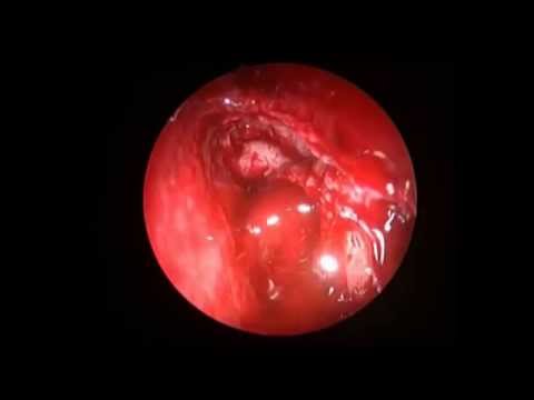
The temporal branch of the facial nerve is a commonly injured nerve during facial trauma due to its superficial course over the zygomatic arch, and is a commonly damaged nerve during facial surgery.1 We report a case of trauma to the left temporal fossa, and subsequent unilateral forehead paralysis. Early exploration revealed external suture compression as the origin of his paralysis. Removal of the suture led to complete resolution of the neurological deficit. The differential diagnosis did not include the possibility of the compression of the nerve by a suture, however the decision for early exploration led to a full recovery.
The anatomy, function, and course of the temporal branch of the facial nerve are critical to the development of a surgical plan. The temporal nerve provides motor innervation to the frontalis, corrugator, and lateral orbicularis oculi muscles.2 These muscles are important for facial expression, and loss of muscle function can have very damaging effects on the patient’s psyche, self-confidence, and social interaction, and can ultimately lead to depression.3 The longer a muscle is denervated, the more the probability of reinnervation decreases, indicating the need for early evaluation, including exploration of the temporal nerve, to expedite the treatment plan and potentially improve outcomes. The risk of the exploratory surgery in certain cases is outweighed by the benefits of returning complete motor function and maintaining normal social interaction.
The decision to provide surgical intervention for the treatment of temporal nerve palsy will be highly dependent on the chance of spontaneous recovery.8 Seventy to eighty percent of patients diagnosed with Bell’s Palsy will have some spontaneous recovery. However, palsy caused by viral infection or trauma will have a lower chance of spontaneous recovery.8 While MRI is only indicated for facial nerve lesions of the brainstem, cerebellopontine angle, and intratemporal locations, EMG may aid in clinical decision making for superficial facial nerve lesions.8 EMG results indicating severe denervation may prompt physicians to either wait for spontaneous improvement or perform exploratory surgery. One study showed that patients with normal EMG results had an 80% chance of full recovery. Patients that showed slight paresis had a 25% chance of full recovery, and patients that lacked excitability on EMG had almost no chance for full recovery.9 Delayed exploration after an EMG indicating slight paresis allows time for the palsy to improve spontaneously, but delay could result in a less favorable outcome in cases of more severe denervation.
More involved procedures may be needed if surgical exploration is delayed or unsuccessful. A primary coaptation of the temporal nerve can be performed with patient outcomes that are aesthetically and functionally pleasing, but this option is most effective in the first 2 months of paralysis.10 The primary repair is effective if both stumps of the nerve are available, the end musculature is still sustainable, and the gap in the nerve is less than 1 centimeter.8,10 When there is a gap of over 1 cm, a graft will need to be harvested to ensure a tension free coaptation.8 After approximately six months to one year of paralysis, the motor end plate of the muscle has a decreased capacity for reanimation, and more drastic repair techniques are indicated.8,11 The temporalis muscle can be transposed locally to reanimate the upper and lower eyelids.8 A free muscle transfer can also be used in muscle reanimation, first using a cross-facial nerve graft and then fixating the neurovascular anastomosis of the transplanted muscle.8,11 However, the results for both procedures are less than optimal. There can be aesthetic changes to the facial area, and the muscles themselves must be retrained to achieve desired function.8 |
| Category |
Education |
| Tags |
Nerve (Anatomical Structure) | Facial Nerve (Cranial Nerve) | Forehead (Anatomical Structure) | Paralysis (Symptom) | Plastic Surgery (Medical Specialty) | mcclellan | McClellan Plastic Surgery | morgantown | West Virginia (US State) |
More Videos