Laparoscopic Repair of Small Umbilical Hernia using Mishra's Knot and Dual Mesh
Hellow guys, Welcome to my website, and you are watching Laparoscopic Repair of Small Umbilical Hernia using Mishra's Knot and Dual Mesh. and this vIdeo is uploaded by Dr. R. K. Mishra at 2024-11-08T07:06:10-08:00. We are pramote this video only for entertainment and educational perpose only. So, I hop you like our website.
Info About This Video
| Name |
Laparoscopic Repair of Small Umbilical Hernia using Mishra's Knot and Dual Mesh |
| Video Uploader |
Video From Dr. R. K. Mishra |
| Upload Date |
This Video Uploaded At 08-11-2024 15:06:10 |
| Video Discription |
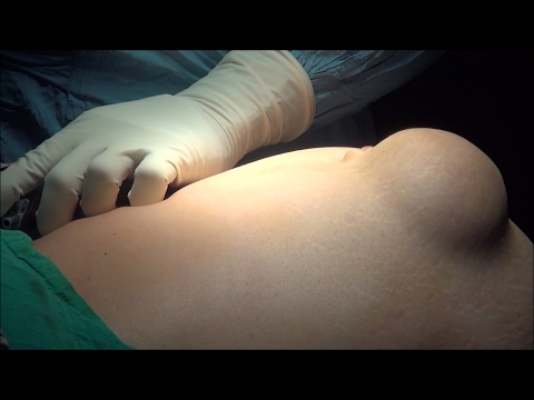
Umbilical hernias are common surgical conditions encountered in adults, often necessitating repair due to the risk of complications such as incarceration or strangulation. With the advent of laparoscopic surgery, minimally invasive techniques have become the gold standard for hernia repairs, offering faster recovery, less postoperative pain, and reduced risk of infection. In this article, we will discuss the laparoscopic repair of a small umbilical hernia using the Mishra’s knot technique and a dual mesh, emphasizing key steps and tips for a successful outcome.
Indications for Laparoscopic Umbilical Hernia Repair
Laparoscopic repair is indicated in patients with symptomatic umbilical hernias, particularly those with recurrent hernias, obese patients, or those seeking faster recovery. Small umbilical hernias, defined as defects less than 3 cm in diameter, can be effectively repaired using a combination of non-absorbable sutures (Mishra's knot) and a dual mesh to reinforce the defect.
Step-by-Step Surgical Technique
1. Initial Access and Creation of Pneumoperitoneum
Mishra's point access is used to introduce a 10 mm trocar under direct vision using the open (Hasson) technique. CO₂ insufflation is initiated to create a pneumoperitoneum with an intra-abdominal pressure of 12-14 mmHg.
A 30° laparoscope is inserted to visualize the hernia defect and surrounding anatomy.
2. Reduction of Hernia Sac
The hernia sac contents, if present, are gently reduced back into the peritoneal cavity using atraumatic graspers.
Ensure complete reduction of any omental or small bowel contents to prevent future complications.
3. Preparation of the Hernia Defect
The edges of the hernia defect are cleaned of any preperitoneal fat. This step is crucial for proper mesh placement and fixation.
Measure the defect size to determine the appropriate size of the dual mesh to be used.
4. Closure of the Defect with Mishra's Knot
The hernia defect is closed using Mishra's knot, a secure extracorporeal knot-tying technique that provides reliable tension and strength. Non-absorbable polypropylene (0) suture is typically used.
The needle is inserted percutaneously using a suture passer, and the defect edges are approximated using the Mishra's knot. This knot has the advantage of being a secure, tension-free closure with minimal risk of knot slippage.
5. Placement of Dual Mesh
A dual mesh (polypropylene side facing the abdominal wall and ePTFE side facing the viscera) is selected to cover the defect with a 3-4 cm overlap on all sides.
The mesh is rolled and introduced through the 10 mm port.
Unroll the mesh within the peritoneal cavity and position it over the defect. The mesh is secured using tacks or absorbable sutures at the periphery to prevent migration.
Ensure there are no folds or wrinkles in the mesh, as this can lead to recurrence or adhesions.
6. Inspection and Closure
Thoroughly inspect the repair for hemostasis and mesh position. The pneumoperitoneum is gradually released, and ports are removed.
Close the supraumbilical port fascia with non-absorbable sutures to prevent port-site hernias.
The skin incisions are closed with absorbable sutures or skin adhesive.
Postoperative Management
Pain Control: Postoperative pain is managed with non-steroidal anti-inflammatory drugs (NSAIDs) and acetaminophen. Opioids are reserved for breakthrough pain.
Mobilization: Early ambulation is encouraged to reduce the risk of deep vein thrombosis (DVT) and promote recovery.
Discharge: Most patients can be discharged on the same day or within 24 hours post-surgery.
Follow-Up: A follow-up appointment is scheduled within one week to check for any signs of infection, seroma, or recurrence.
Advantages of Using Mishra's Knot and Dual Mesh
Secure Closure: The use of Mishra's knot provides a secure closure of the hernia defect, reducing the risk of recurrence.
Dual Mesh: The combination of polypropylene and ePTFE materials in the dual mesh provides strength while minimizing the risk of adhesions to the bowel.
Minimally Invasive Approach: Laparoscopic repair offers the advantages of smaller incisions, less postoperative pain, and faster recovery compared to open repair.
Tips and Tricks for a Successful Repair
Ensure adequate overlap of the mesh over the defect to minimize the risk of recurrence.
Mishra's knot technique requires practice to achieve optimal tension; however, it provides a robust, tension-free closure once mastered.
Avoid excessive use of tacks near critical structures such as bowel or blood vessels to prevent postoperative complications.
Potential Complications
Seroma Formation: Can occur but typically resolves spontaneously. Aspiration is rarely required.
Mesh Infection: Rare but requires prompt management, including antibiotics or, in severe cases, mesh removal.
Recurrence: Unlikely with proper technique and adequate mesh overlap. |
| Category |
Education |
| Tags |
Laparoscopic Hernia Repair | Umbilical Hernia | Mishra's Knot | Dual Mesh | Minimally Invasive Surgery | Laparoscopic Technique | Hernia Defect Closure | Extracorporeal Knot | Hernia Surgery | Surgical Mesh | Laparoscopic Suturing | Hernia Recurrence | Surgical Tips | Umbilical Hernia Repair | Mesh Fixation | Hernia Repair Complications | Postoperative Recovery | Minimally Invasive Hernia Repair | Laparoscopic Port Placement |
More Videos