The Cystohepatic Mesentery: A new Anatomical Surgical Perspective To The Triangle of Calot
Hellow guys, Welcome to my website, and you are watching The Cystohepatic Mesentery: A new Anatomical Surgical Perspective To The Triangle of Calot. and this vIdeo is uploaded by Prof. Abdelghany Elshamy at 2022-03-06T09:20:05-08:00. We are pramote this video only for entertainment and educational perpose only. So, I hop you like our website.
Info About This Video
| Name |
The Cystohepatic Mesentery: A new Anatomical Surgical Perspective To The Triangle of Calot |
| Video Uploader |
Video From Prof. Abdelghany Elshamy |
| Upload Date |
This Video Uploaded At 06-03-2022 17:20:05 |
| Video Discription |
Laparoscopic Cholecystectomy : The Cystohepatic mesentery and Cystohepatic Space and the Critical View of Safety - our Laparoscopic Anatomical Surgical Perspective.
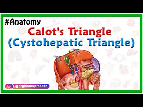
This is our Current laparoscopic anatomical surgical perspective of the CYSTOHEPATIC TRIANGLE or TRIANGLE OF CALOT .
During laparoscopic cholecystectomy as seen in the video, dissection of this triangle (or i think is better to be termed The CYSTOHEPATIC MESENTERY (CHM) or CYSTOHEPATIC MESENTERIC SPACE (CHMS), we have observed that this small mesentery is formed of an anterior visceral peitoneal layer , then the fatty layer in the middle , then the posterior peritoneal layer . So , it is formed of three layers (trilaminar), in which the fatty layer is invested between the anterior and posterior visceral peitoneal layers.
The contents of this mesentery include the cystic artery or better to refer to it as cystic blood vessels (as there are frequently anterior and posterior branches or sometimes multiple small vessels) , the cystic lymph node , and other minute lymphatics and nerve fibres.
The cystic artery gives off multiple small branches that supply the cystic duct crossing in the cystohepatic mesentery (nearly in a ladder-steps shaped configuration as seen in the video), these small vascular branches to cystic duct forms a plexus of vascular network around the cystic duct. So, we have to put this into consideration during dissection around the cystic duct, to avoid troublesome bleeding from the cystic artery and avoiding injury of surrounding structures.
The Laparoscopic or Endoscopic Surgical implications :
During laparoscopic cholecystectomy we usually start dissection in this space anteriorly by dissection of the anterior peitoneal layer to visualize the cystic duct, and cystic artery or cystic vessels. Then the middle fatty layer is visualized and dissected , then the posterior layer is dissected . I prefer to dissect the posterior layer after reflecting the neck of the gallbladder or the Hartmann's pouch anteriorly and medially and dissect it better from posterior under direct vision or it can be dissected from anterior after dissection of the fatty layer.
Most laparoscopic surgeons have observed that the cystic lymph node of Lund is considered as a landmark of the cystic artery or its anterior branch near the neck of the gallbladder.
During dissection of the posterior layer of CYSTOHEPATIC MESENTERY we have to be causious to avoid bleeding from a posterior branch of cystic artery or bleeding from right hepatic artery with a caterpillar course or accessory right hepatic artery . Usually we complement the anterior dissection with dissection of the posterior peritoneal layer from posterior to visualize anomalous course or accessory right hepatic or large posterior cystic vessel to avoid bleeding.
The cystic lymph node may be small and sometimes large according to the inflammation, it may be partially dissected and retracted inferomedially towards the common hepatic duct just to visualize the cystic artery to deal with it near to the gallbladder and away from the common hepatic duct as this is more safe avoiding injury to the bile duct or bleeding in this space , this lymph node may somtimes be removed to visualize the cystic artery, but I prefer to preserve it as possible .
Impacted stones or Mirizzi syndrome will lead to deformed or partial or near total obliteration of Calot triangle or CYSTOHEPATIC TRIANGLE and that is why it is better to be termed CYSTOHEPATIC MESENTERY or CYSTOHEPATIC MESENTERIC SPACE or CYSTOHEPATIC SPACE.
Also , acute and chronic inflammation will lead to inflammation in the CYSTOHEPATIC MESENTERY and it Will be shortened and thickened and deformed making the space smaller or even about to be obliterated with difficulty in dissection and identification of structures due to fibrosis, excessive vascularity and thickening and shotening of Cystohepatic Mesentery.
The dissection should be away from the Common hepatic duct and the common bile duct should not be dissected except in cases of CBD exploration or for proper visualization before ligation or clipping of cystic duct.
The Value of this detailed laparoscopic or microscopic Surgical Anatomy :
Our Aim is to achieve the critical view of safety with Safe Cholecystectomy in the era of laparoscopic surgery. This can be achieved with current understanding of detailed laparoscopic anatomy of the Cystohepatic triangle OR Cystohepatic Mesentery.
This will facilitate identification of the structures with great precision avoiding injury of common hepatic duct, Common bile duct and, anomalous right hepatic artery and bleeding with its consequences in this space and important area during laparoscopic cholecystectomy .
These anatomical consideration can be applied to open cholecystectomy as well. |
| Category |
People & Blogs |
| Tags |
People & Blogs Download MP4 | People & Blogs Download MP3 | People & Blogs Download MP4 360p | People & Blogs Download MP4 480p | People & Blogs Download MP4 720p | People & Blogs Download MP4 1080p |
More Videos