Left endometriotic mass with parametrial nodule invading the left uterine horn (Part 4/5)
Hellow guys, Welcome to my website, and you are watching Left endometriotic mass with parametrial nodule invading the left uterine horn (Part 4/5). and this vIdeo is uploaded by Safe Laparoscopy at 2019-01-20T06:00:03-08:00. We are pramote this video only for entertainment and educational perpose only. So, I hop you like our website.
Info About This Video
| Name |
Left endometriotic mass with parametrial nodule invading the left uterine horn (Part 4/5) |
| Video Uploader |
Video From Safe Laparoscopy |
| Upload Date |
This Video Uploaded At 20-01-2019 14:00:03 |
| Video Discription |
Tips and Tricks │Step by Step technique│Safe Laparoscopy
Subscribe to Our Channel https://bit.ly/2AwCggo
Procedure
Ureterolysis, adnexaectomy with hysteretomy and metroplasty
This represents Part 4 of our "Progressive learning section on how to resect deep endometriotic nodules"
Case
36 year old nulliparous patient with severe dysmenorrhea.
She had undergone laparoscopic surgery twice to remove a left a ovarian endometrioma
Pre op investigations
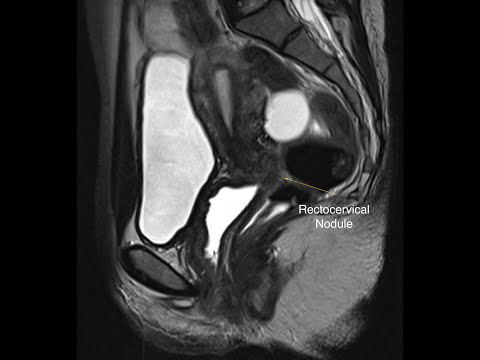
Vaginal ultrasound: Left tubo-ovarian mass occupying the left pelvis and left parametrium. The right ovary appeared normal. The uterus showed signs of left myometrial thickening in conjunction with the turbo-ovarian mass
Examination
Vaginal assessment showed a very tender large left nodule lateral, as well as involving, the left utero-sacral ligament
Radiology
An IVU examination was organized, which showed bilateral normal ureters without distention
The situation was discussed with the patient. Despite her nulliparous status, It was decided to proceed with laparoscopy and most likely left adnexaectomy
After introducing the laparoscope, a thorough assessment of all abdomino-pelvic organs was carried out
The uterus was normal as well as the right adnexa. However, there was a left tube-ovarian mass, firmly adherent to the left pelvic sidewall, left parametrium and invading the left uterine horn.
It was decided as planned to proceed with left adnexaectomy.
Step 1
The thin bowel was cleared form the pelvis and the sigmoid colon was detached from the abdominal sidewall and the peritoneum opened. The left ureter was identified.
Step 2
The peritoneum lateral to the left adnexa was opened all the way to the pelvic inlet.
The left round ligament was desiccated and cut with bipolar current
Step 3
The adnexal mass was mobilized medially and displaced from the large pelvic vessels. The left ureter was identified again and dissected free from its surrounding structures whilst maintaining its vascular support.
Step 4
The ureter was further mobilized and freed
Step 5
The infundibulo-pelvic ligament was identified, freed, desiccated and cut
Step 6
The superior vescical as well as left uterine artery was identified, desiccated and cut. Thus the mass and the ureter were further mobilized.
It became obvious that there was a large endometriotic nodule under the left ureter extending to the left uterosacral ligament
Step 7
The ureter was dissected free from the surrounding mass all the way close to the bladder
Step 8
The turbo ovarian mass was lifted and the nodule dissected with part of the left utero-sacral ligament with the use of bipolar scissors
Step 9
Once the mass was reasonably mobilized, vasopressin was injected into the uterine wall and the mass resected with a monopolar hook. A portion of the left horn as well as a small area of the uterine wall were removed
Step 10
The uterine wound was closed with monocril 0 sutures
Step 11
Hemostasis was assessed and the the pelvic and abdominal cavities repeatedly washed
Step 12
Final view of the left pelvic retro-peritoneal anatomy shown.
Conclusive thoughts
When operating on deep endometriosis, it is vital to know our retro-peritoneal anatomy as intra-peritoneal resection of such lesions, is often impossible
As mentioned before haemostasis is vital in order to recognize structures. Speed is not an ally in such cases. Slow meticulous surgery is required to avoid accidental damage to the large vessels, and the ureter.
It is important to resect the disease completely otherwise pain will recur. In this case, complete removal would not have been possible without partial metrectomy. The transection of the uterine vessels, in this case, was of no consequence to the uterine physiology.
Surgical team
G. Pistofidis
S. Kogeorgos
P. Balinakos
K. Dimitropoulos
ΤΙΤΛΟΣ: ΛΑΠΑΡΟΣΚΟΠΙΚΗ ΑΦΑΙΡΕΣΗ ΟΖΟΥ ΕΝΔΟΜΗΤΡΙΩΣΗΣ ΑΡ ΠΑΡΑΜΗΤΡΙΟΥ - ΑΡ ΟΥΡΗΤΗΡΑ Ο ΟΠΟΙΟΣ ΔΙΗΘΕΙ ΤΟ ΑΡ ΚΕΡΑΣ ΤΗΣ ΜΗΤΡΑΣ│ ΧΕΙΡΟΥΡΓΙΚΟΙ ΧΡΟΝΟΙ ΒΗΜΑ ΠΡΟΣ ΒΗΜΑ │ΜΙΑ ΑΣΦΑΛΗΣ ΤΕΧΝΙΚΗ
ΧΕΙΡΟΥΡΓΙΚΗ ΟΜΑΔΑ - ΛΕΥΚΟΣ ΣΤΑΥΡΟΣ ΑΘΗΝΩΝ:
ΠΙΣΤΟΦΙΔΗΣ Γ.
ΚΟΓΕΩΡΓΟΣ Σ.
ΜΠΑΛΙΝΑΚΟΣ Π.
ΔΗΜΗΤΡΟΠΟΥΛΟΣ Κ. |
| Category |
Science & Technology |
| Tags |
yt:quality=high | endometriotic mass | parametrial nodule invading the left ureter | laparoscopy | Laparoscopy | Adenomyosis | Pelvic pain:laparoscopy | Endometriosis treatment | Safe laparoscopy | endometriosis treatment | laparoscopic surgery | laproscopy operation | stage 4 endometriosis | vagina | endometriosis vlog | surgery | what is endometriosis | endometriosis and pregnancy | infertility | nucleus medical media | abdomen | procedure | ovarian | lap for endo | endometriosis | medical | endometriosis surgery |
More Videos